

The debate surrounding myofibrillar hypertrophy versus sarcoplasmic growth has persisted in exercise science for decades, often oversimplified into binary categories that fail to capture the nuanced biology occurring within skeletal muscle fibers. At our laboratory, we have committed significant analytical resources to dissecting the mechanistic underpinnings of each hypertrophic pathway—not as competing phenomena, but as concurrent adaptations whose relative magnitudes are governed by training variables, training status, and the molecular signaling environment within the myocyte. In this comprehensive analysis, we present the current state of evidence, clarify persistent misconceptions, and offer precise operational definitions that practitioners can use to inform programming decisions.
Before we can evaluate training implications, we must establish unambiguous definitions. The terms “myofibrillar hypertrophy” and “sarcoplasmic hypertrophy” describe distinct structural changes within a muscle fiber, and conflating them—or treating them as mere theoretical constructs—undermines evidence-based programming.
Myofibrillar Hypertrophy: Contractile Protein Accretion
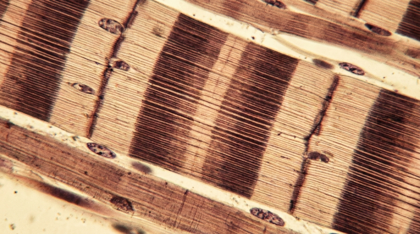
Myofibrillar hypertrophy refers to the addition of contractile proteins (actin and myosin) within existing myofibrils and/or the parallel addition of new myofibrils within a muscle fiber. This process directly increases the force-generating capacity of the fiber because more cross-bridge cycling sites are available per unit of cross-sectional area. The functional outcome is an increase in specific tension—the force produced per unit of muscle cross-sectional area—assuming the contractile protein density increases relative to total fiber volume.
We define a related concept, myofibrillar packing, as the condition in which myofibrils grow faster than the surrounding sarcoplasmic volume. In this scenario, the ratio of contractile protein to total intracellular content shifts upward, potentially increasing specific tension without necessarily expanding overall fiber cross-section proportionally. One rodent study has hinted at slight myofibrillar packing under high-load conditions, though we must note that translating murine data directly to human physiology requires caution given differences in fiber-type distribution and remodeling kinetics.
Sarcoplasmic Hypertrophy: Non-Contractile Volume Expansion
Sarcoplasmic hypertrophy refers to the expansion of the non-contractile, fluid-rich compartment of the muscle fiber, which includes the sarcoplasm, sarcoplasmic reticulum, mitochondria, glycogen stores, water, and critically—sarcoplasmic proteins such as glycolytic enzymes, creatine kinase, and other metabolic machinery. For years, critics dismissed sarcoplasmic hypertrophy as a myth or reduced it to mere glycogen supercompensation. We now have definitive evidence that this is incorrect.
Research published as recently as January 2026 confirms that high-volume weightlifting promotes sarcoplasmic hypertrophy in young men, increasing muscle fluid and metabolic proteins with measurably less strength gain per unit of muscle size compared to high-load, low-volume protocols. Crucially, the sarcoplasmic protein fraction—not just glycogen and water—drives this expansion. Data from elite-level lifters demonstrates approximately 10% lower myofibril density compared to recreationally trained individuals, despite possessing substantially larger muscle cross-sectional areas. This finding is incompatible with a model in which all hypertrophy is purely myofibrillar.
In exploring the intricacies of muscle growth, a related article that delves into the mechanisms of hypertrophy is available at this link: The Science of Myofibrillar Hypertrophy vs. Sarcoplasmic Growth. This article provides valuable insights into the differences between myofibrillar and sarcoplasmic hypertrophy, helping readers understand how various training techniques can influence muscle development and overall performance. By examining these two types of growth, fitness enthusiasts can better tailor their workout routines to achieve specific goals.
Mechanistic Drivers: What Triggers Each Pathway
Understanding the molecular signaling cascades that preferentially activate one pathway over the other allows us to move beyond correlational observation and toward causal programming logic.
Mechanical Tension and the mTORC1 Axis
High mechanical tension—generated through heavy loading at or above approximately 70-85% of one-repetition maximum—activates the mechanotransduction pathway involving focal adhesion kinase (FAK), phosphatidic acid (PA), and ultimately the mammalian target of rapamycin complex 1 (mTORC1). This signaling cascade preferentially upregulates ribosomal biogenesis and the translation of contractile protein mRNA. The primary downstream outcome is the synthesis of actin and myosin at rates that exceed sarcoplasmic protein synthesis, favoring myofibrillar hypertrophy.
We observe that high-load, low-volume protocols—typically characterized by sets of 3-6 repetitions with loads exceeding 80% 1RM—maximize this mechanical tension signal while minimizing metabolic byproduct accumulation. The January 2026 data corroborates this framework: subjects performing high-load, low-volume training demonstrated functional strength gains that were disproportionately large relative to cross-sectional area increases, consistent with myofibrillar packing or at minimum proportional myofibrillar growth.
Metabolic Stress and Sarcoplasmic Protein Upregulation
Conversely, protocols that maximize time under tension and metabolic byproduct accumulation—hydrogen ions, inorganic phosphate, lactate, and reactive oxygen species—activate a partially distinct adaptive response. The cellular environment created by high-volume, moderate-load training (typically 8-15+ repetitions per set with shorter rest intervals) demands enhanced metabolic buffering capacity, glycolytic enzyme density, and sarcoplasmic reticulum calcium handling efficiency.
The adaptive response to this stimulus is the preferential synthesis of sarcoplasmic proteins—glycolytic enzymes, heat shock proteins, and metabolic intermediaries—that expand the sarcoplasmic compartment relative to the myofibrillar fraction. This is not non-functional growth; it represents a legitimate adaptation to the metabolic demands imposed. However, it does not contribute directly to maximal force production, which is why high-volume trainees may exhibit larger muscles with lower pound-for-pound strength metrics.
The Role of Training Status in Pathway Dominance
We must emphasize a critical finding from human studies: in untrained individuals undergoing 6-12 weeks of resistance training, myofibril growth appears to be proportional to overall fiber growth. That is, the ratio of contractile protein to total fiber volume remains relatively constant during the initial adaptation phase. Sarcoplasmic dominance does not appear to manifest in novice trainees over short training durations.
This proportional growth pattern suggests that the divergence between myofibrillar and sarcoplasmic hypertrophy is a phenomenon that emerges with training advancement—likely requiring years of accumulated training volume to produce measurable dissociations. The 10% myofibril density deficit observed in elite lifters versus less-trained individuals represents the cumulative product of thousands of training sessions, not a rapid acute response.
Fiber-Type Considerations and Specificity
The hypertrophic pathway activated is not independent of the fiber population being recruited. We must account for the heterogeneous composition of human skeletal muscle.
Type I Fibers and Sarcoplasmic Adaptation
Type I (slow-oxidative) fibers possess inherently larger sarcoplasmic volumes, more mitochondria, and greater capillary density. These fibers are preferentially recruited during lower-intensity, higher-duration contractions. Training protocols that maximize Type I fiber recruitment and metabolic stress may disproportionately expand the sarcoplasmic compartment simply because the targeted fiber population already has a lower myofibril-to-sarcoplasm ratio.
Type II Fibers and Myofibrillar Dominance
Type II (fast-glycolytic) fibers, particularly Type IIx and Type IIa subtypes, possess higher myofibrillar density and greater force-generating capacity per unit area. High-load training preferentially recruits these fibers via Henneman’s size principle (high-threshold motor unit recruitment). The combination of high mechanical tension and Type II fiber targeting creates the optimal signaling environment for myofibrillar protein accretion.
Fiber-Type Transitions as a Confounding Variable
We note that chronic resistance training induces fiber-type transitions—predominantly from Type IIx toward Type IIa. This shift itself alters the sarcoplasmic-to-myofibrillar ratio within converted fibers and must be accounted for when interpreting biopsy data. Researchers who fail to control for fiber-type composition changes may misattribute structural shifts to one hypertrophic pathway when the underlying cause is a transition in the fiber population itself.
Sure, here is the sentence with the clickable link:
Check out the latest fitness program at Hypertrophy Protocol for effective muscle building.
Advanced Mechanisms Beyond the Binary Framework
The myofibrillar-versus-sarcoplasmic dichotomy, while useful as an organizing framework, is insufficient to capture the full complexity of muscle adaptation in advanced trainees.
Myonuclear Accretion and the Myonuclear Domain Theory
Each muscle fiber contains multiple nuclei (myonuclei), and each nucleus governs protein synthesis within a finite cytoplasmic volume—its “myonuclear domain.” When a fiber expands beyond the synthetic capacity of existing myonuclei, satellite cells must fuse with the fiber to donate additional nuclei. This myonuclear accretion process is a rate-limiting step in sustained hypertrophy and operates independently of whether the growth is myofibrillar or sarcoplasmic.
We observe that protocols which maximize mechanical damage and inflammatory signaling—eccentric-heavy loading, novel movement patterns—most effectively activate satellite cell proliferation and fusion. This suggests that periodization strategies incorporating controlled muscle damage phases may facilitate long-term growth of both types by expanding the myonuclear pool.
Ribosomal Biogenesis as a Growth Ceiling
The translational capacity of a muscle fiber—determined by ribosome density—establishes an upper limit on protein synthesis rates. Advanced trainees who have maximized ribosomal content may experience diminished hypertrophic responses regardless of training stimulus quality. Emerging evidence suggests that deload periods or strategic training cessation may allow ribosomal pools to regenerate, potentially explaining the “supercompensation” effect observed after planned recovery phases.
Connective Tissue and Extracellular Matrix Remodeling
We also note that not all “muscle size” gains occur within the muscle fiber itself. The extracellular matrix (ECM), including the endomysium and perimysium, remodels in response to training and contributes to overall limb circumference and imaging-based measures of muscle cross-sectional area. This ECM remodeling is neither myofibrillar nor sarcoplasmic hypertrophy—it is a third category often overlooked in the binary debate.
In exploring the intricacies of muscle growth, the article on The Science of Myofibrillar Hypertrophy vs. Sarcoplasmic Growth offers valuable insights into the different mechanisms that contribute to strength and size. For those interested in further understanding how these processes can be optimized for better training results, a related article on hypertrophy training techniques can be found at Hypertrophy Protocol. This resource delves into practical strategies that can enhance your workout regimen and maximize muscle development.
Practical Programming Implications
| Metrics | Myofibrillar Hypertrophy | Sarcoplasmic Growth |
|---|---|---|
| Primary Focus | Increasing the size and strength of muscle fibers | Increasing the volume of fluid and energy stores within the muscle |
| Effect on Muscle Density | Increases muscle density | May not significantly increase muscle density |
| Effect on Muscle Endurance | May improve muscle endurance | Can improve muscle endurance due to increased energy stores |
| Training Approach | Focuses on heavy lifting with lower reps | Involves higher rep ranges and shorter rest periods |
| Appearance | Results in a more defined and sculpted look | May result in a more “pumped” appearance |
Based on our analysis of the current evidence base, we offer the following programming considerations for practitioners seeking to preferentially target one hypertrophic pathway.
For Myofibrillar Hypertrophy Prioritization
- Load selection: 80-90% of 1RM, sets of 2-6 repetitions
- Rest intervals: 3-5 minutes to allow full phosphocreatine resynthesis and neural recovery
- Volume: Moderate total sets (12-16 per muscle group per week), emphasizing intensity over accumulated fatigue
- Tempo: Controlled eccentrics (3-4 seconds) with maximal concentric intent
- Expected outcome: Disproportionate strength gains relative to size increases; improved specific tension
For Sarcoplasmic Hypertrophy Prioritization
- Load selection: 60-75% of 1RM, sets of 8-15+ repetitions
- Rest intervals: 60-90 seconds to maintain metabolic stress accumulation
- Volume: High total sets (16-25+ per muscle group per week), emphasizing accumulated work and metabolic byproduct generation
- Tempo: Continuous tension with minimal lockout; prolonged time under tension per set (40-70 seconds)
- Expected outcome: Greater visual size increases with moderate strength gains; increased muscular endurance
Integrated Periodization for Complete Development
We recommend that most trainees employ undulating or block periodization that alternates between mechanical tension-dominant and metabolic stress-dominant phases. This approach ensures that both myofibrillar and sarcoplasmic compartments receive adequate adaptive stimuli across the training macrocycle, prevents accommodation to either stimulus type, and supports the myonuclear accretion and ribosomal biogenesis processes that underpin long-term growth capacity.
In exploring the intricacies of muscle growth, a fascinating article that complements the discussion on myofibrillar hypertrophy versus sarcoplasmic growth can be found at this link. It delves into the various training methodologies that can influence these distinct types of hypertrophy, providing valuable insights for those looking to optimize their workout routines. Understanding the differences between these growth mechanisms can significantly enhance one’s approach to strength training and overall fitness.
Conclusions and Future Research Directions
The evidence base as of early 2026 confirms that sarcoplasmic hypertrophy is a real, measurable phenomenon driven by sarcoplasmic protein increases—not merely glycogen or water retention. Simultaneously, myofibrillar hypertrophy remains the primary driver of functional strength development and is preferentially activated by high-load mechanical tension protocols. The divergence between these pathways appears to manifest most clearly in advanced trainees over extended training timelines, while novice trainees experience proportional growth during initial adaptation periods.
Our key institutional position is this: the myofibrillar versus sarcoplasmic framework is valid and practically useful, but it must be understood as a continuum of relative emphasis rather than a binary switch. All resistance training produces some degree of both adaptations; programming variables modulate the ratio, not the absolute presence or absence of either pathway.
Future research priorities for our laboratory include longitudinal biopsy studies in intermediate-to-advanced trainees exceeding 12-week durations, single-fiber proteomic analyses to quantify sarcoplasmic protein species changes with training, and the interaction between nutritional periodization (particularly carbohydrate and creatine availability) and sarcoplasmic volume regulation. We anticipate that the next five years will yield substantially more granular data on these mechanisms, enabling increasingly precise programming recommendations for athletes seeking specific hypertrophic outcomes.