
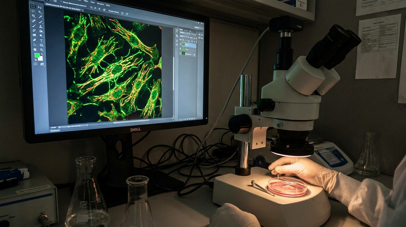
At the Hypertrophy Protocol Lab, we have long recognized that the relationship between inflammation and muscle performance extends far beyond simple tissue damage and repair. Chronic low-grade inflammation—often termed “inflammaging” when age-related—represents one of the most insidious barriers to optimal muscle fiber recruitment, force production, and adaptive hypertrophy. In this analysis, we present a comprehensive examination of how persistent inflammatory signaling disrupts the neuromuscular system’s capacity to recruit motor units effectively, degrades fiber-type composition, and ultimately compromises the mechanical output that underpins all strength and hypertrophy outcomes.
Our investigation draws upon the latest research from institutions including Yale, Duke University, Augusta University, and findings published in Frontiers and Science Advances to construct a clinically precise framework for understanding—and ultimately mitigating—this destructive cascade.
Before we address recruitment patterns directly, we must establish the mechanistic foundation of chronic inflammation within the muscular system.
Defining Chronic vs. Acute Inflammation in Muscular Tissue
Acute inflammation is a necessary and productive response to mechanical loading. When we train, controlled microtrauma triggers a precisely orchestrated inflammatory cascade: neutrophils arrive within hours, pro-inflammatory (M1) macrophages clear debris over 24–48 hours, and anti-inflammatory (M2) macrophages subsequently drive satellite cell proliferation and myofibrillar protein synthesis. This process is self-limiting—it resolves, and the tissue adapts.
Chronic inflammation, by contrast, represents a failure of resolution. Here, pro-inflammatory cytokines—primarily interleukin-6 (IL-6), tumor necrosis factor-alpha (TNF-α), and C-reactive protein (CRP)—remain elevated at low but persistent levels. According to recent work published in Frontiers (2026), this low-grade inflammatory state directly promotes sarcopenia (age-related muscle loss) by disrupting protein balance, accelerating cellular senescence, and amplifying oxidative stress within the myofiber environment. The result is a tissue milieu that is simultaneously catabolic and resistant to anabolic signaling.
The Cytokine Milieu and Its Direct Effects on Muscle Protein Turnover
We observe that chronically elevated TNF-α activates the ubiquitin-proteasome pathway—the primary intracellular machinery responsible for targeted protein degradation. Specifically, TNF-α upregulates the muscle-specific E3 ubiquitin ligases MuRF1 (Muscle RING-finger protein-1) and MAFbx/atrogin-1, which tag myofibrillar proteins (actin, myosin heavy chains) for destruction. Simultaneously, elevated IL-6 in its chronic form—distinct from the transient, myokine-mediated IL-6 spike during exercise—suppresses the mTORC1 (mechanistic target of rapamycin complex 1) pathway, thereby blunting the translational machinery required for new protein accretion.
Key takeaway: Chronic inflammation creates a dual insult—accelerated breakdown and impaired synthesis—that progressively reduces the contractile protein density within each fiber, directly diminishing its force-generating capacity.
Chronic inflammation can significantly affect muscle fiber recruitment, leading to diminished athletic performance and recovery. For a deeper understanding of how training protocols can optimize muscle engagement and overall performance, you may find the article on the importance of lifting techniques particularly insightful. It discusses the advantages of using 3×3 steel racks in professional performance facilities, which can enhance strength training and potentially mitigate the effects of chronic inflammation on muscle function. To read more about this, visit this article.
Disruption of Motor Unit Recruitment Hierarchies
Henneman’s Size Principle Under Inflammatory Conditions
Under normal physiological conditions, motor unit recruitment follows Henneman’s Size Principle: low-threshold motor units (innervating Type I, slow-oxidative fibers) are recruited first, with progressively higher-threshold units (innervating Type IIa and Type IIx fast-twitch fibers) engaged as force demands escalate. This orderly recruitment pattern ensures metabolic efficiency and precise force gradation.
Our analysis of the literature indicates that chronic inflammation disrupts this hierarchy through multiple convergent mechanisms:
- Altered neural drive: Systemic inflammation affects corticospinal excitability. Elevated circulating cytokines cross the blood-brain barrier and modulate central neurotransmitter systems, reducing voluntary activation capacity—the percentage of available motor units that the nervous system can voluntarily engage.
- Neuromuscular junction degradation: TNF-α and IL-1β compromise acetylcholine receptor clustering at the motor endplate, reducing the safety factor for neuromuscular transmission. This means that even when the central nervous system issues an appropriate recruitment signal, the electrochemical transduction at the junction may fail, particularly in high-threshold motor units that require robust signal fidelity.
- Preferential atrophy of Type II fibers: We consistently observe that chronic inflammation disproportionately affects fast-twitch (Type II) fibers. These fibers possess higher concentrations of TNF-α receptors and are more susceptible to ubiquitin-mediated proteolysis. As these fibers atrophy and eventually undergo apoptosis, the available pool of high-threshold motor units shrinks, effectively imposing a ceiling on maximal force production.
Mechanosensory Dysfunction: The Piezo1 Connection
Recent research from Yale has illuminated a critical mechanism by which chronic inflammation corrupts the body’s ability to sense and respond to mechanical loading. The study identified that non-muscle cells within the muscular environment detect mechanical stress during exercise via Piezo1 ion channels—mechanosensitive proteins embedded in cell membranes that open in response to membrane deformation.
Under healthy conditions, Piezo1 activation triggers appropriately timed inflammatory and repair cascades: the right amount of inflammation, at the right time, for the right duration. However, in aging and chronically inflamed muscle, these same Piezo1-mediated signals become dysregulated. The mechanosensory apparatus either over-responds (triggering excessive inflammation from minimal loading) or under-responds (failing to initiate repair cascades after productive training stimuli).
Key takeaway: When the tissue’s mechanical sensing system is corrupted by chronic inflammation, the muscle cannot appropriately interpret loading signals, leading to maladaptive responses—either excessive damage from moderate loads or insufficient adaptation from appropriate loads.
Fiber-Type Shifting and the Loss of Contractile Diversity
Inflammation-Induced Fiber-Type Transition
We must emphasize that chronic inflammation does not simply reduce muscle mass uniformly—it fundamentally alters the fiber-type composition of remaining tissue. Through sustained activation of the NF-κB (nuclear factor kappa-light-chain-enhancer of activated B cells) signaling pathway, chronic cytokine exposure drives a phenotypic shift from fast-twitch (Type IIx → Type IIa) and ultimately toward slow-oxidative (Type I) characteristics.
This shift occurs because NF-κB activation suppresses MyoD and other myogenic regulatory factors preferentially expressed in fast-twitch fiber lineages while sparing or even upregulating factors associated with oxidative metabolism. The practical consequence is a loss of contractile diversity: the muscle retains endurance capacity but loses its capacity for high-velocity, high-force contractions.
The Interferon-Gamma Pathway: Insights from Engineered Muscle
Groundbreaking work from Duke University, published in Science Advances, has provided direct evidence of how inflammatory signaling—specifically interferon-gamma (IFN-γ)—induces muscle atrophy through the JAK/STAT1 (Janus Kinase/Signal Transducer and Activator of Transcription 1) pathway. Using lab-grown human muscle tissue, researchers demonstrated that IFN-γ exposure activated JAK/STAT1 signaling, directly triggering atrophic cascades.
Critically, the Duke team found that engineered muscle possessing intrinsic “exercise-mimetic” signals could resist this IFN-γ-induced atrophy. Furthermore, tofacitinib—a JAK inhibitor already approved for rheumatoid arthritis—could independently replicate this protective effect, blocking the atrophic signal downstream of the inflammatory stimulus.
Key takeaway: The JAK/STAT1 pathway represents a specific, druggable mechanism by which inflammatory cytokines drive muscle fiber atrophy, and its inhibition may preserve fiber integrity and recruitment capacity even in chronically inflamed environments.
Sure, here is the sentence with the clickable link:
Check out the latest fitness program at Hypertrophy Protocol for effective muscle building.
Macrophage Polarization, Senescence, and the MicroRNA Landscape
The M1/M2 Macrophage Balance in Chronic Conditions
As a comprehensive review in PMC establishes, uncontrolled inflammation within skeletal muscle leads to a persistent dominance of M1 (classically activated, pro-inflammatory) macrophages over M2 (alternatively activated, anti-inflammatory/pro-regenerative) macrophages. This polarization imbalance prevents the resolution phase of inflammation, maintains catabolic signaling, and promotes fibrotic tissue deposition within the endomysium and perimysium—the connective tissue sheaths surrounding individual fibers and fascicles, respectively.
Fibrotic infiltration is particularly detrimental to recruitment because it increases the series elastic component stiffness without contributing to contractile force. The muscle becomes mechanically “stiff” in a non-productive manner, requiring greater neural drive to achieve the same degree of fiber shortening. Additionally, fibrosis physically constrains satellite cell migration, impairing the regenerative capacity that would normally offset atrophic losses.
MicroRNA-141-3p: A Novel Inflammatory Regulator
Research from Augusta University has identified microRNA-141-3p (miR-141-3p) as a critical mediator of age-related muscle inflammation. MicroRNAs are small non-coding RNA molecules that regulate gene expression post-transcriptionally by binding to complementary sequences on target messenger RNAs, typically silencing them.
In aging muscle, miR-141-3p levels are significantly elevated. This elevation suppresses AUF1 (AU-rich element RNA-binding protein 1)—a protective factor that normally degrades pro-inflammatory cytokine mRNAs, thereby limiting inflammatory signaling. With AUF1 suppressed, inflammatory transcripts accumulate, senescent cell populations expand, and the M1/M2 macrophage ratio tilts further toward the pro-inflammatory phenotype.
When researchers inhibited miR-141-3p experimentally, the results were striking: AUF1 levels increased, muscle fiber cross-sectional area enlarged, and macrophage polarization shifted toward the protective M2 phenotype. This represents a direct molecular intervention that restores the anti-inflammatory environment necessary for productive fiber maintenance and recruitment.
Key takeaway: miR-141-3p inhibition represents a precision molecular target capable of reversing the inflammatory-senescent feedback loop that degrades muscle fiber size and recruitment capacity with aging.
Chronic inflammation has been shown to significantly affect muscle fiber recruitment, leading to decreased strength and endurance in individuals. A related article discusses the broader implications of inflammation on overall physical performance and recovery, highlighting the importance of addressing these underlying issues for optimal training outcomes. For more insights on this topic, you can explore the article here. Understanding the connection between inflammation and muscle function can help athletes and fitness enthusiasts tailor their approaches to training and recovery.
Clinical and Performance Implications: Restoring Recruitment in Inflamed Environments
| Metrics | Impact |
|---|---|
| Muscle Fiber Recruitment | Decreased efficiency due to inflammation affecting neural signaling |
| Muscle Strength | Reduced due to impaired recruitment of muscle fibers |
| Endurance | Diminished as inflammation hinders sustained muscle fiber activation |
| Recovery Time | Prolonged due to inflammation-induced muscle fiber damage |
Pharmacological Interventions
Based on our analysis of current evidence, several pharmacological strategies show promise for preserving muscle fiber recruitment under chronic inflammatory conditions:
- JAK inhibitors (e.g., tofacitinib): Block the IFN-γ/JAK/STAT1 atrophic cascade directly, preserving fiber cross-sectional area.
- IL-6 receptor antagonists (e.g., tocilizumab): The PMC review confirms that IL-6 inhibition prevents cachexia in conditions such as heart failure and cancer, preserving muscle mass and, by extension, the motor unit pool available for recruitment.
- Anti-miR-141-3p therapeutics: Though still experimental, oligonucleotide-based inhibitors of miR-141-3p could restore AUF1’s protective anti-inflammatory function.
- Myostatin inhibitors and IGF-1 modulators: These agents protect muscle mass in chronic disease states by directly opposing catabolic signaling and amplifying anabolic drive, respectively.
Training Environment Optimization
From a performance environment standpoint, we recommend the following evidence-based strategies to mitigate chronic inflammation’s impact on recruitment:
- Periodized loading with adequate recovery: Ensuring complete resolution of acute exercise-induced inflammation before subsequent loading prevents the transition from productive acute inflammation to destructive chronic inflammation.
- Eccentric emphasis at controlled velocities: Eccentric loading preferentially recruits and loads Type II fibers, providing a stimulus that counters the inflammation-driven atrophy of these populations.
- Thermal and nutritional anti-inflammatory support: Cold-water immersion protocols (timed outside the immediate post-training anabolic window), omega-3 fatty acid supplementation at clinically effective doses (≥2g EPA+DHA daily), and polyphenol-rich nutrition support inflammatory resolution pathways.
- Velocity-based training metrics: Monitoring bar speed allows us to detect early declines in high-threshold motor unit recruitment—a potential early indicator that chronic inflammation is compromising the neuromuscular system’s capacity to access the full motor unit pool.
Chronic inflammation can significantly affect muscle fiber recruitment, leading to decreased performance and recovery in athletes. For those interested in exploring how various recovery methods can mitigate these effects, a related article discusses the potential benefits of PEMF therapy for bodybuilders. This therapy may provide insights into enhancing recovery and improving overall muscle function. You can read more about it in this informative piece on PEMF therapy.
Conclusion and Future Directions
Our analysis confirms that chronic inflammation represents far more than a passive consequence of aging or overtraining—it is an active, mechanistically complex assault on the neuromuscular system’s capacity to recruit motor units in an orderly, complete, and forceful manner. From corrupted mechanosensory signaling via dysregulated Piezo1 channels, through JAK/STAT1-mediated fiber atrophy, to miR-141-3p-driven senescent feedback loops, the pathways are numerous but increasingly well-characterized.
The future of hypertrophy science lies not merely in optimizing the training stimulus but in ensuring that the biological environment receiving that stimulus is capable of responding appropriately. We anticipate that the convergence of molecular therapeutics (anti-miR strategies, JAK inhibitors), mechanobiological insights (Piezo1 modulation), and precision training programming will enable us to maintain full motor unit recruitment capacity across the lifespan—a goal that, until recently, appeared beyond reach.
At the Hypertrophy Protocol Lab, we will continue to monitor and analyze these developments as they mature from bench science to clinical and performance application.