

Within our laboratory, we have observed a growing body of clinical evidence suggesting that red light therapy—more precisely termed photobiomodulation (PBM)—offers neurological advantages that extend well beyond superficial tissue repair. For strength athletes operating under high neuromuscular demand, repeated mechanical stress, and significant central nervous system (CNS) fatigue, these benefits represent a meaningful frontier in performance environment optimization.
In the following analysis, we present our assessment of the current evidence base, focusing on the mechanisms through which PBM interacts with neural tissue, the implications for strength sport populations, and the practical parameters that govern effective application.
Before we examine neurological outcomes, we must establish the biophysical foundation of red light therapy. Photobiomodulation refers to the application of light—typically in the red (620–700 nm) and near-infrared (NIR, 700–1100 nm) spectral ranges—to biological tissue at non-thermal intensities. The primary chromophore (light-absorbing molecule) responsible for the downstream cascade is cytochrome c oxidase (CCO), located on Complex IV of the mitochondrial electron transport chain.
Mitochondrial Energetics and ATP Production
When photons at appropriate wavelengths are absorbed by CCO, we observe a dissociation of nitric oxide (NO) from the enzyme’s binuclear center. This dissociation restores oxygen binding capacity, accelerates electron transport, and increases the proton gradient across the inner mitochondrial membrane. The net result is enhanced adenosine triphosphate (ATP) synthesis—the cell’s fundamental energy currency.
For neural tissue, which is among the most metabolically demanding in the body, this upregulation of mitochondrial output carries significant functional implications. Neurons operating under energetic stress—whether from repeated high-threshold motor unit recruitment during maximal lifting or from accumulated microtrauma—benefit directly from restored oxidative phosphorylation capacity.
Reactive Oxygen Species Modulation and Cellular Signaling
We note that PBM does not merely increase ATP. At appropriate doses (typically 10–50 joules per treatment zone), the therapy produces a transient, low-level increase in reactive oxygen species (ROS) that activates transcription factors such as NF-κB and AP-1. These factors upregulate genes involved in anti-inflammatory cascades, antioxidant defense, and neurotrophin production—including brain-derived neurotrophic factor (BDNF), which we will discuss in subsequent sections.
Key Takeaway: PBM operates through mitochondrial photon absorption, enhancing ATP output and triggering cytoprotective signaling cascades that are directly relevant to neural tissue under athletic stress.
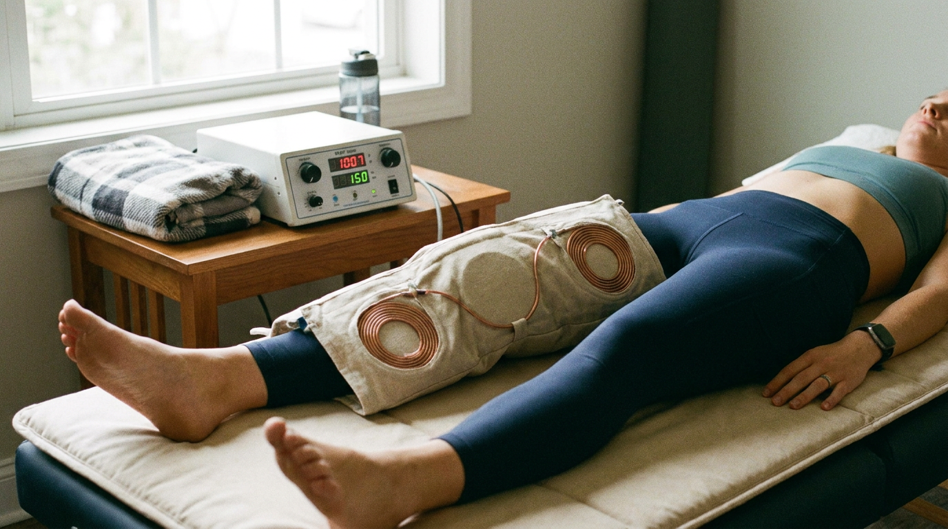
In exploring the neurological benefits of red light therapy for strength athletes, it’s interesting to consider complementary recovery methods that may enhance performance and recovery. A related article discusses the potential of PEMF therapy for bodybuilders, examining whether this technology can effectively aid in recovery and muscle performance. For more insights on this topic, you can read the article here: PEMF Therapy for Bodybuilders: Does It Actually Work?.
Neuroprotection Against Repetitive Impact and Neuroinflammation
One of the most compelling recent findings relevant to strength athletes—particularly those in contact-intensive training environments such as strongman, rugby-supplemented strength programs, or combat sport crossover athletes—comes from a January 2026 preliminary trial conducted at the University of Utah.
The University of Utah Football Trial (2026)
In this study, 26 college football players were exposed to transcranial photobiomodulation (tPBM) using a Vielight Neuro Gamma headset delivering 810 nm near-infrared light pulsed at 40 Hz (gamma frequency). Over a 16-week competitive season, researchers monitored neuroinflammatory biomarkers and brain microstructure via advanced imaging.
The findings were notable: the treatment group showed no significant increase in neuroinflammation over the season, while the placebo group exhibited the expected inflammatory escalation associated with repetitive head acceleration events (RHAE). Brain microstructure—assessed via diffusion tensor imaging metrics—remained stable in the PBM cohort, suggesting preservation of white matter integrity.
Implications for Strength Athletes
We recognize that the immediate association between football and strength sports may seem tangential. However, we must account for several realities within strength training environments:
- Heavy axial loading during squats, deadlifts, and overhead presses transmits force through the cervical spine and cranium
- Missed lifts and jarring impacts during strongman events (atlas stones, log press catches) create acute cranial acceleration
- Accumulated sub-concussive exposure in athletes who cross-train in wrestling, martial arts, or contact sports
For these populations, the mechanism demonstrated in the Utah trial—enhanced mitochondrial function, improved cerebral blood flow, and reduced neuroinflammatory signaling—represents a viable prophylactic strategy against long-term neural degradation. Larger trials involving 300 veterans and first responders are currently underway to validate these findings at scale.
Key Takeaway: Transcranial PBM at 810 nm/40 Hz pulsing prevented neuroinflammatory escalation over a 16-week season of repetitive impacts, preserving brain microstructure in ways directly applicable to strength athletes with cranial loading exposure.
CNS Recovery and Neuromuscular Performance Optimization
Strength performance is not purely a muscular phenomenon. We consistently emphasize within our protocols that maximal force production is governed by the CNS’s capacity to recruit high-threshold motor units, maintain firing rate, and sustain descending drive from motor cortical areas. When the CNS is fatigued—a state we term central fatigue—peripheral muscle capacity becomes irrelevant because the neural signal driving contraction is degraded.
Central Fatigue Mechanisms in Strength Athletes
Central fatigue accumulates through multiple pathways:
- Serotonergic upregulation in brainstem raphe nuclei, reducing motor cortex excitability
- Depletion of prefrontal dopaminergic drive, reducing motivation and voluntary activation capacity
- Neuroinflammatory cytokine elevation (IL-6, TNF-α) crossing the blood-brain barrier post-training
- Reduced cerebral oxygenation during prolonged high-effort sessions
PBM addresses several of these pathways simultaneously. By enhancing mitochondrial function in cortical neurons, increasing local cerebral blood flow via nitric oxide-mediated vasodilation, and attenuating neuroinflammatory signaling, transcranial red light therapy creates conditions favorable for accelerated CNS recovery between training sessions.
Practical Neuromuscular Implications
We interpret the available evidence to suggest that strength athletes applying tPBM post-session or on recovery days may experience:
- Faster restoration of voluntary activation capacity (the percentage of available motor units an athlete can consciously recruit)
- Improved inter-session readiness for high-intensity neural work (singles, doubles, triples at >90% 1RM)
- Reduced perception of effort at submaximal loads during subsequent sessions
While direct maximal voluntary contraction (MVC) increases have not been demonstrated in current meta-analyses, the facilitation of recovery enables higher training frequency and accumulated volume—which are the actual drivers of long-term strength adaptation.
Key Takeaway: PBM targets central fatigue pathways by restoring cortical mitochondrial function and reducing neuroinflammation, enabling faster CNS recovery and higher-quality subsequent training sessions.
Sure, here is the sentence with the clickable link:
Check out the latest fitness program at Hypertrophy Protocol for effective muscle building.
Cognitive Function, Sleep Architecture, and Training Quality
We cannot separate neurological health from cognitive performance, nor can we isolate either from sleep quality. For strength athletes, these domains converge in practical ways that directly influence training outcomes.
Cognitive Preservation Under Chronic Training Stress
Prior research (spanning 8–10 week tPBM protocols) has demonstrated improved cognitive function in adults exposed to repetitive head acceleration events. Specifically, improvements in executive function, working memory, and reaction time were documented. For strength athletes, these cognitive domains translate to:
- Improved proprioceptive awareness during complex lifts (reducing injury risk)
- Enhanced motor learning during technique acquisition phases
- Better intra-set decision-making (recognizing when to terminate a set approaching failure)
Sleep Architecture Optimization
We observe that 40 Hz gamma-frequency pulsed NIR light—the same parameter used in the Utah trial—coincides with endogenous gamma oscillation frequencies associated with memory consolidation and deep sleep transitions. Emerging evidence suggests that tPBM applied in evening protocols may support:
- Faster sleep onset latency
- Increased slow-wave sleep (SWS) duration, during which growth hormone secretion peaks
- Improved sleep continuity, reducing nighttime cortisol spikes that impair recovery
For the strength athlete, sleep is not merely rest—it is the primary anabolic window. Any intervention that reliably improves sleep architecture has downstream effects on protein synthesis rates, hormonal milieu, and next-day training capacity.
Mood and Motivational Drive
We note anecdotal and preliminary clinical observations that consistent tPBM use correlates with improved mood stability and reduced training-related anxiety. While we await larger controlled trials, the proposed mechanism—enhanced prefrontal cortical metabolism and improved serotonin-dopamine balance—is biologically plausible and consistent with PBM’s known effects on mitochondrial function in frontal lobe tissue.
Key Takeaway: PBM’s neurological benefits extend beyond acute recovery to include preserved cognitive function, optimized sleep architecture, and stabilized mood—all of which compound into superior long-term training quality.
In exploring the various recovery methods available to strength athletes, one intriguing approach is the application of red light therapy, which has been shown to offer significant neurological benefits. For those interested in understanding how mechanical tension plays a crucial role in muscle growth and recovery, a related article delves into this topic in detail. You can read more about it in this insightful piece on the role of mechanical tension in modern hypertrophy protocols. This connection highlights the importance of combining different recovery strategies to optimize performance and enhance overall athletic outcomes.
Peripheral Neurological Benefits: Muscle Recovery and Afferent Signaling
| Neurological Benefits of Red Light Therapy for Strength Athletes |
|---|
| Improved motor function |
| Enhanced muscle coordination |
| Increased reaction time |
| Reduced muscle fatigue |
| Enhanced cognitive function |
While our primary focus has been on central neurological effects, we must address the peripheral nervous system implications supported by robust 2024–2025 meta-analytic data.
Reduction in Muscle Damage Biomarkers
Multiple systematic reviews have confirmed that pre-exercise PBM application (10–50 J per muscle group) significantly reduces:
- Creatine kinase (CK) levels at 24–96 hours post-exercise (indicating less sarcolemmal disruption)
- Delayed-onset muscle soreness (DOMS) severity
- Lactate dehydrogenase (LDH) elevation
Neural Implications of Reduced Peripheral Damage
We emphasize that muscle damage is not merely a local tissue event. Damaged muscle fibers release damage-associated molecular patterns (DAMPs) that activate peripheral nociceptors—sensory neurons transmitting pain signals. This nociceptive barrage:
- Sensitizes spinal cord dorsal horn neurons (central sensitization)
- Inhibits motor neuron output via protective reflex arcs (arthrokinetic inhibition)
- Alters movement patterning as the CNS compensates around perceived threat
By reducing the magnitude of peripheral damage, PBM indirectly preserves normal motor neuron recruitment patterns and movement quality in the days following high-eccentric-load sessions. This is particularly relevant for strength athletes performing heavy negatives, deficit movements, or high-volume hypertrophy blocks.
Fatigue Delay and Endurance of Neural Drive
Meta-analytic data also demonstrate that pre-exercise PBM delays fatigue onset during sustained contractions. We interpret this partially as a peripheral metabolic effect (improved mitochondrial efficiency in muscle fibers), but also as a reduced rate of afferent fatigue signaling—meaning the CNS receives fewer inhibitory signals from type III/IV muscle afferents, allowing sustained motor output for longer durations.
Key Takeaway: Pre-exercise PBM at 10–50 J reduces peripheral damage markers and nociceptive signaling, preserving motor recruitment quality and delaying central fatigue accumulation during training.
Practical Application Parameters and Protocol Considerations
We conclude with our current evidence-based recommendations for strength athletes seeking to integrate PBM into their neurological recovery protocols.
Wavelength Selection
- 810 nm (near-infrared): Optimal for transcranial application; sufficient penetration depth to reach cortical tissue through scalp and skull
- 630–670 nm (red): Appropriate for superficial peripheral applications (muscle tissue, superficial nerve beds)
Dosimetry
- Transcranial protocols: 10–25 J/cm² delivered over 20–25 minutes; pulsed at 40 Hz for gamma-frequency entrainment
- Peripheral/muscle protocols: 10–50 J per muscle group; applied pre-exercise for protective effects or post-exercise for recovery acceleration
Timing
- Pre-training (peripheral): 5–30 minutes before session onset for fatigue delay and damage mitigation
- Post-training (transcranial): Within 2 hours of session completion for CNS recovery initiation
- Evening (transcranial): 20-minute session 60–90 minutes before sleep for architecture optimization
Equipment Considerations
We note that the clinical trial evidence referenced in this analysis utilized medical-grade devices (specifically, the Vielight Neuro Gamma for transcranial applications). Consumer-grade panels vary substantially in irradiance, wavelength accuracy, and pulsing capability. We recommend athletes verify:
- Independently measured irradiance (mW/cm² at treatment distance)
- Wavelength certification within ±5 nm of target
- Pulsing frequency accuracy (if using gamma-entrainment protocols)
Key Takeaway: Effective neurological PBM requires precise wavelength (810 nm transcranial), appropriate dosimetry (10–50 J), and timing aligned with training demands. Device quality verification is non-negotiable for clinical-grade outcomes.
Summary Position
We regard photobiomodulation as one of the more scientifically substantiated recovery modalities available to strength athletes—particularly when evaluated through the lens of neurological preservation and CNS recovery optimization. The convergence of neuroprotective evidence (University of Utah, 2026), peripheral recovery meta-analyses (2024–2025), and cognitive-sleep literature positions PBM as a modality warranting serious integration into high-performance strength training environments.
We will continue monitoring the larger-scale trials currently underway and will update our protocols as dose-response relationships and long-term outcome data become available. For now, the evidence supports a clear conclusion: the brain and nervous system of the strength athlete deserve the same deliberate recovery investment as the musculoskeletal system, and photobiomodulation represents a viable, evidence-informed tool for that purpose.