
Last reviewed October 20, 2026. This guide is based on published research, and every number below comes from the study linked beside it.
Sarcoplasmic hypertrophy means a muscle fiber grows mainly by expanding the fluid, enzymes and other material around its contractile machinery, rather than adding contractile protein in proportion. It can show up in humans, most clearly when trained lifters suddenly jump to very high, unaccustomed volumes. Most studies, though, find that the contractile part of the fiber grows roughly in step with the fiber, and the best recent imaging work points to new myofibrils as the main driver of fiber growth.
It shouldn’t change how you train. No study shows you can choose “sarcoplasmic” or “myofibrillar” growth by picking a rep range, and light and heavy loads build similar amounts of muscle when sets are taken to failure. The familiar line that bodybuilders build puffy sarcoplasmic muscle while powerlifters build dense myofibrillar muscle rests on a few small cross-sectional studies that conflict with others.
At a glance
| Verdict | It can appear, mainly after sudden very high volume. Most fiber growth comes from new myofibrils. |
|---|---|
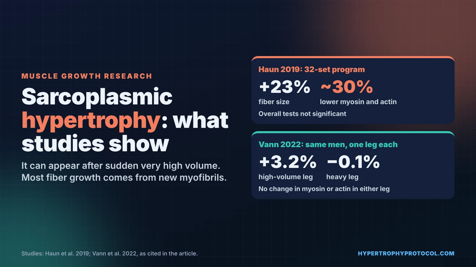
| Effect size / key numbers | Haun 2019: fibers +23%, myosin and actin about 30% lower after a 32-set program; overall tests not significant. |
| Evidence strength | Weak and conflicting: small biopsy studies, and bodybuilder-vs-powerlifter data are cross-sectional only. |
| What to do | Don’t train for it. Take sets close to failure, progress load or reps, pick rep ranges for practical reasons. |
| Main caveat | A biopsy samples a tiny piece of muscle, and swelling can look like sarcoplasmic growth. |
Research last reviewed October 20, 2026. See the sections below for sources.
What is sarcoplasmic hypertrophy, exactly?
A muscle fiber is mostly packed with myofibrils, the bundles of actin and myosin that produce force. The space around them, called the sarcoplasm, holds water, glycogen, mitochondria, enzymes and other proteins. A 2020 review from the lab that has studied this most estimated that myofibrils occupy about 85% of the space inside a fiber, and that muscle tissue is about 75% water, 10 to 15% myofibrillar protein and about 5% sarcoplasmic protein (Roberts et al., 2020).
The review describes three ways a fiber could grow:
- Conventional hypertrophy. Myofibrils and sarcoplasm expand in proportion. If a fiber’s cross-section grows 20% and myofibrils fill 85% of it, about 17 of those 20 points would be new myofibril and about 3 would be sarcoplasm.
- Sarcoplasmic hypertrophy. The sarcoplasm expands faster than the myofibrils, so the fiber gets bigger while its contractile protein becomes less concentrated.
- Myofibril packing. Myofibrils are added faster than the fiber grows, so the fiber becomes more densely packed with contractile protein.
Myofibrillar hypertrophy, in the way lifters use the term, is the first and third patterns: adding contractile protein. The scientific question isn’t whether sarcoplasm exists or grows. It’s whether it grows out of proportion to the myofibrils, how often, and whether that matters.
Why is sarcoplasmic hypertrophy so hard to measure?
Every method needs a muscle biopsy, and each one has a blind spot. Roberts and colleagues list four:
| Method | What it measures | Main weakness |
|---|---|---|
| Electron microscopy | Area taken up by myofibrils, mitochondria and the space between them | Uses 1 to 2 mg of tissue; 20 images cover parts of only 20 to 40 cells; processing can distort spacing |
| Phalloidin staining | Actin signal inside each fiber outline | Also binds non-contractile actin; poor freezing leaves gaps that look like empty space |
| Gel electrophoresis (SDS-PAGE) | Myosin and actin per milligram of muscle | A drop per milligram can mean swelling rather than new sarcoplasm; leftover blood in a sample inflates its weight |
| Single-fiber specific tension | Force per unit of fiber area, an indirect sign of contractile density | Often fewer than 10 fibers per person; preparation makes fibers swell |
Sampling is the deeper problem. A vastus lateralis biopsy yields about 100 mg of tissue, from a muscle the review estimates weighs more than 500 g in young, healthy adults. The review puts that at 1/2,500th of the muscle (100 mg of a 500 g muscle actually works out to 1/5,000th), and the pre- and post-training biopsies can’t come from the same bundle of fibers. So when a study reports that myosin per milligram fell 30%, that’s a real measurement, but it’s a long way from proof that a whole muscle is “puffier.”
What did Haun et al. 2019 actually find?
This is the study most often cited as proof of sarcoplasmic hypertrophy (Haun et al., 2019). Thirty trained young men, with about five years of lifting behind them, followed six weeks of very high-volume training, three days a week. Volume ramped from 10 to 32 sets per week for each exercise, so the final week included 32 sets of 10 back squats.
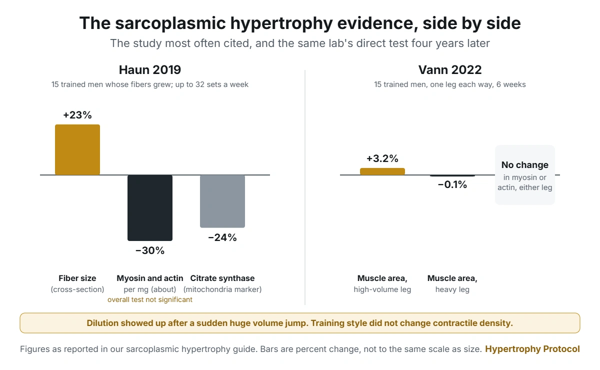
The researchers analyzed the 15 men whose fibers grew. Their average fiber cross-section rose 23%, yet myosin and actin concentrations fell about 30%, and citrate synthase activity, a marker of mitochondrial content, fell 24%. Forty sarcoplasmic proteins rose, including enzymes involved in glycolysis. Seven of the men gave another biopsy eight days after training stopped: sarcoplasmic protein was still 66% above baseline, and actin and myosin were still about 50% lower.
That’s a measured result, and it’s the strongest human evidence that sarcoplasmic expansion can happen. It comes with real limits:
- The week-6 biopsy came 24 hours after the last session of an extremely hard program, so swelling from muscle damage is a plausible confounder. The authors argued against it because tissue fluid didn’t rise, but the same lab’s later review noted that body-wide extracellular fluid did rise and contributed to measured muscle mass gains in this cohort.
- Only half the participants grew. The other half saw no change or a decrease in fiber size.
- The overall statistical tests for myosin and actin weren’t significant (p = 0.052 and p = 0.055). The roughly 30% drops came from follow-up comparisons the authors ran anyway.
- A 2026 review added that one participant started with unusually high actin and myosin values, and that removing him made the decline non-significant (Van Every et al., 2026).
- Participants were split across whey protein and maltodextrin supplement groups, and there was no lower-volume or heavy-load comparison group.
What happened when the same lab tested heavier or more normal training?
The follow-up studies are where the “high reps make sarcoplasmic muscle” idea starts to fall apart.
In 15 trained men who did 10 weeks of low-volume, heavy training at 68 to 90% of their max, type II fibers grew, while sarcoplasmic protein concentration and tissue fluid didn’t change (Vann et al., 2020). Myosin trended down 2.9% and actin fell 3.2%. Even the heavy-load program produced a slight dilution of contractile protein, not the “denser” muscle the myth predicts.
The most direct test came next. Fifteen trained men trained one leg with higher volume and lighter loads and the other with heavier loads, three times a week for six weeks (Vann et al., 2022). The high-volume leg grew 3.2% in cross-sectional area by MRI, while the heavy leg changed by −0.1%. The heavy leg gained more leg extensor strength. Neither leg showed any change in myosin or actin abundance, which the authors said challenged their own earlier findings. The high-volume leg did show higher non-myofibrillar protein synthesis over the six weeks.
Studies in beginners point the same way. In 15 untrained men, 10 weeks of twice-weekly training increased fiber size while the share of fiber area occupied by myofibrils stayed the same, which suggests the myofibrils expanded in proportion (Ruple et al., 2021). The area occupied by mitochondria did rise. The Roberts review also summarizes a 12-week study in untrained men in which the 13 best responders grew their fibers 34% with no change in myosin or actin per milligram.
The newest structural evidence comes from a microscopy method that can count and size individual myofibrils in fiber cross-sections. Using mouse and human models of increased loading, the researchers found that fiber growth was largely driven by myofibrillogenesis, the creation of new myofibrils (Jorgenson et al., 2024). That’s growth of the contractile machinery itself, the opposite of the sarcoplasmic story.
Do bodybuilders really have “puffy” muscle and powerlifters “dense” muscle?
This is the claim most people have heard, and the evidence is mixed. The best-known support comes from single-fiber testing in 12 bodybuilders, 6 power athletes and 14 untrained controls (Meijer et al., 2015).
- Bodybuilders’ fibers were 67% larger than the power athletes’ and 88% larger than the controls’.
- Their fibers produced 32% more total force than controls, and the power athletes’ produced 50% more.
- Force per unit of fiber area was 62% lower in bodybuilders than in power athletes and 41% lower than in controls.
The authors suggested lower myofibrillar density or changes to the contractile proteins as possible causes, and noted that part of the difference may simply come from fibers being very large. Bodybuilder fibers were still stronger than untrained fibers in absolute terms.
Other comparisons point the other way. The Roberts review summarizes studies in which well-trained lifters had specific tension similar to untrained men, one in which type II specific tension was higher in well-trained bodybuilders than in recreational lifters, and one in which trained men had about 9% more myosin and actin per milligram of muscle than untrained men. Older electron microscopy work found experienced lifters had about 30% less space filled by myofibrils and twice the sarcoplasmic space of untrained men. The review’s own conclusion is that these discordant findings make it hard to say how years of training affect myofibril density.
These are all cross-sectional snapshots of small groups. They can’t separate training style from genetics, years of training, body size or anything else that differs between a bodybuilder and a powerlifter.
Some of the “fullness” lifters notice is glycogen and water, which training in general increases. The 2026 review notes that each gram of glycogen binds 3 to 4 grams of water, that bodybuilders have been reported to carry about double the glycogen of sedentary people, and that intracellular water rose 8.2% in men and 11.0% in women over a 16-week bodybuilding-style program. That’s normal training adaptation, not a separate, inferior kind of muscle.
What the gym claims say vs what studies measured
| Claim | What was actually measured | Verdict |
|---|---|---|
| High reps build sarcoplasmic muscle; heavy weights build myofibrillar muscle | Same men, one leg each style: no change in myosin or actin in either leg (Vann 2022) | Not supported |
| Bodybuilders’ muscle is less dense than powerlifters’ | Lower force per fiber area in 12 bodybuilders (Meijer 2015); similar or higher in other small groups | Mixed; cross-sectional only |
| Sarcoplasmic hypertrophy doesn’t exist | Contractile protein diluted and sarcoplasmic proteins rose after a 32-set program (Haun 2019) | Too strong; it can appear, mainly after sudden very high volume |
| Sarcoplasmic growth is just water | Glycolytic enzymes and other sarcoplasmic proteins increased (Haun 2019); fluid shifts also contributed | Partly; it’s protein and fluid |
| Chasing the pump builds sarcoplasmic size | 2026 review: claims that cell swelling or the pump meaningfully drive hypertrophy aren’t supported by evidence | Not supported |
| Most training growth is myofibrillar | Proportional myofibril expansion in beginners (Ruple 2021); new myofibrils drive fiber growth (Jorgenson 2024) | Best supported |
Does lifting light weights build a different kind of muscle?
Not in any way studies have detected. A meta-analysis of 21 studies compared light loads, at or below 60% of max, with heavier loads, with every set taken to failure (Schoenfeld et al., 2017). Muscle growth was similar between conditions. One-rep-max strength improved more with heavy loads, while isometric strength didn’t differ.
That strength gap is the root of the myth. Heavy lifters test better on a heavy single, so people assume their muscle must be built differently. But a one-rep max is a skill as well as a measure of muscle, and heavy training is much closer practice for it. The within-subject 2022 trial shows the same pattern at the cellular level: the heavy leg got stronger and the high-volume leg grew more, with no difference in contractile protein density between them.
The 2026 review goes further, concluding that sarcoplasmic hypertrophy as a distinct, functionally meaningful contributor to growth lacks strong evidence, and proposing that it isn’t a meaningful contributor to hypertrophy. Researchers who study it still disagree on how often it happens. But when the lab that reported it most clearly tested high-volume and heavy training in the same men, contractile protein density didn’t differ between the two.
Should sarcoplasmic hypertrophy change how you train?
No. Train the way the broader hypertrophy evidence supports, and the sarcoplasm will take care of itself.
- Take sets close to failure. Hard sets that put real load on the muscle are what grow it. Mechanical tension and muscle growth explains why.
- Pick rep ranges for practical reasons. Anywhere you can reach near failure with good form will build muscle. Lift heavy if you also want a bigger one-rep max, since that strength is partly a skill you have to practice.
- Add load or reps over time. Progressive overload in a small home gym covers how to keep progressing with limited equipment.
- Choose exercises for tension and range. Training at long muscle lengths and machines vs free weights matter far more than any sarcoplasmic-versus-myofibrillar label.
- Be wary of sudden huge volume jumps. The clearest sarcoplasmic signal came after trained men more than tripled their weekly sets in six weeks. Some of a quick size gain from a big volume jump may be fluid and diluted contractile protein, so judge progress over months.
- Eat enough protein. New myofibrils are built from it. The protein calculator gives a daily target and a per-meal split.
Frequently asked questions
Is sarcoplasmic hypertrophy real?
It has been measured in some human studies, most clearly after very high, unaccustomed training volume. Whether it’s a lasting adaptation, a short-term phase of growth or partly swelling is unresolved. Most training studies find contractile protein grows roughly in proportion to fiber size.
What is the difference between sarcoplasmic and myofibrillar hypertrophy?
Myofibrillar hypertrophy is growth from added contractile protein, the actin and myosin that produce force. Sarcoplasmic hypertrophy is growth in which the surrounding fluid, enzymes and other material expand faster than the contractile protein. In practice, normal training produces both, mostly in proportion.
Does sarcoplasmic size disappear faster when you stop training?
Nobody has measured that directly. The Roberts review describes a study in which fiber size rose 16% over 90 days of training, then returned to pre-training levels within about 10 days of stopping. The authors speculated that shrinking sarcoplasm could explain such a fast drop, but they didn’t test it.
Are bodybuilders weaker than they look?
Per unit of fiber area, bodybuilders’ fibers produced 41% less force than untrained fibers in one study of 12 bodybuilders. Their much larger fibers still produced 32% more total force than untrained fibers. Other small studies found trained lifters’ fibers were just as dense as untrained ones.
Related guides
- Mechanical Tension and Muscle Growth: What Drives Hypertrophy
- Progressive Overload in a Small Home Gym
- Training at Long Muscle Lengths: What the Evidence Supports
- Machines vs Free Weights for Muscle Growth: The Research
- Protein Calculator: daily target and per-meal split
- Weekly Sets Planner: count your hard sets per muscle
- All free Hypertrophy Protocol tools